
Before some surgeons at Indiana University Health begin a complex operation, they turn to a 500-pound machine in IU Health’s 3D printing studio for a consult.
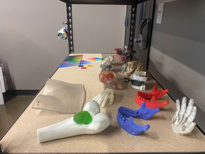
Five machines, nestled in the single-room studio in a technology park on the west side of Indianapolis, produce models of patients’ anatomy, from hearts to spines to jaws to brains, allowing surgeons to develop better roadmaps for the procedures they perform. These models can also help surgeons counsel patients before they reach the operating room.

“Just like an athlete would think about a game or practice before they do it, these models allow us to do the same thing,” said IU Health orthopedic surgeon Dr. Christopher Collier, who has used three models produced by the studio.
Since the lab opened about a year ago, it has fulfilled 92 3D-printing orders from clinicians, according to clinical applications specialist Kara Lutes, one of the Ricoh contract workers who staffs the lab. Those orders include patient-specific models, anatomic models for educational or research use and even objects like cupholders or glasses frames to use in occupational therapy.
On a recent afternoon, Hannah Cahill, a Ricoh senior clinical engineer, demonstrated the process, creating a model of a child’s hand and wrist in one of the studio’s five printers. The machine works like an inkjet printer, that is, if that inkjet printer extruded filaments of plastic, cured them with ultraviolet light and flattened them into perfectly level layers with a miniature rolling pin.
Twenty hours later, the machine spits out a model of a child’s forearm. Cahill removes excess material with a water jet and the model is ready for use.
This model is a replica of one that Cahill produced for an IU Health surgical team that was planning to operate on a child with Madelung’s deformity, a congenital condition that causes one arm bone, the radius, to grow abnormally. The surgeons planned to shorten the child’s ulna to realign both arm bones at the wrist.
With the original model in hand, the physicians were able to explain the procedure to the child and family before operating.
Cahill says she produces roughly one model organ for IU Health physicians each week.
Each model is based on MRI or CT scans of the patient. Cahill uses software to transform these scans into printable models. Depending on a model’s size and complexity, the printing process can take anywhere from 30 minutes to four days.
That results in a turnaround time of roughly one week, said Collier, who has used three models made in the studio in recent months. His team had used 3D models for the past several years, but those models came from third-party vendors in a process that generally took weeks and could add between $5,000 and $20,000 to a patient’s medical bills.
Printing the models in-house is faster and cheaper, he said.
Earlier this year, Collier’s team was preparing to operate on a child with rhabdomyosarcoma. The child’s cancer wasn’t particularly aggressive, but a tumor had twisted around his spine, causing scoliosis that could impact his mobility and breathing. Because the anatomy was so complicated – sections of the child’s spine were nearly horizontal – Collier commissioned a model of the spine and the tumor.
“Our entire surgical team before the surgery was able to hold onto this model in our hands,” Collier said. That helped them create a playbook for how to access the tumor, remove it, and straighten the child’s spine, down to calculating the length of screws and rods needed to secure it in place.
Collier said he often gives the 3D-printed anatomical models to his patients once their operations are done.
“They’ve found that to be really impactful too – just as a memento of what they’ve had to overcome,” he said.
IU Health surgeon Avinash Mantravadi, who specializes in head and neck oncology and reconstructive surgery, uses 3D-printed models of patients’ organs to adjust surgical plates to fit their unique anatomy days before they check into the hospital. This saves both time and money, he said in a video posted to IU Health’s Instagram.
Cardiothoracic surgeon Dr. Jeremy Herrmann uses models from the 3D printing studio to help make his surgical plans and to prevent any surprises in the operating room.
“These give us a clearer picture of what we’re going to encounter when we actually get in surgery,” he said in an interview June 25, as he held up a model of the upper chambers of a patient’s heart. “If I can look at this and hold it and turn it – and think about the relationships of all these different structures, and where we might be able to cut into something or reconnect something – that probably makes the whole surgery go faster.”
Research backs up the idea that 3D-printed organs are big time-savers. Across seven studies, using 3D-printed anatomical models shortened operations by 62 minutes on average, according to a paper published in Academic Radiology in 2020.
Collier said he hopes the studio will encourage physicians and researchers to continue to innovate.
“When things are close and in-house and easy, people are going to come up with new ways to use this technology,” he said.
Tilly Robinson is a Pulliam fellow for the Indianapolis Star. She can be reached at tilly.robinson@indystar.com.
This article originally appeared on Indianapolis Star: IU Health is 3D-printing model organs to make surgeries faster
Reporting by Tilly Robinson, Indianapolis Star / Indianapolis Star
USA TODAY Network via Reuters Connect
By Tilly Robinson, Indianapolis Star | USA TODAY Network